MULTIDISCIPLINARY TEAM
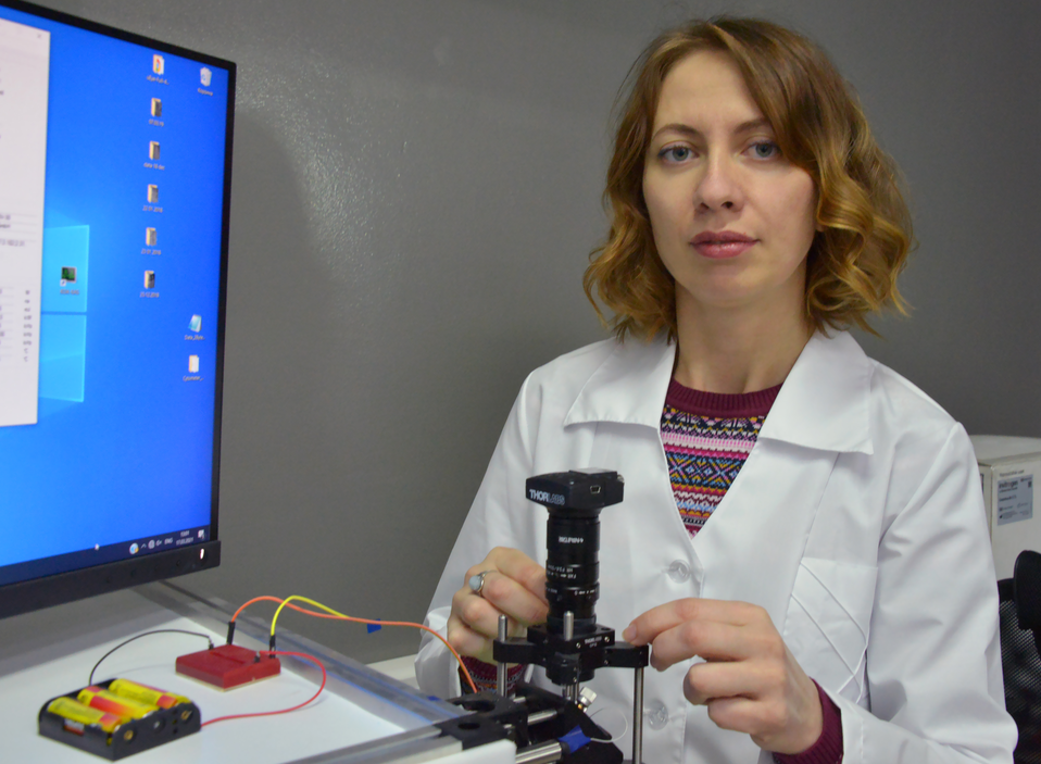
An experimental sample of a multifunctional diagnostic test system based on a spectral microscopy platform, created by young scientists of Saratov State University, can make it possible to detect tumour cells at the earliest stage of the disease. It's all about ‘magic’ or simply innovative blood clearing. The research is carried out in the Laboratory of Biomedical Photoacoustics, SSU, and supported by the Ministry of Science and Higher Education of the Russian Federation within the framework of the Science national project.
It is known that the majority of deaths (up to 90%) are caused by the spread of cancer cells from the primary tumour through the circulatory system to various organs, where they form secondary tumours called metastases. Scientists have shown that spectroscopic methods in combination with optical clarification make it possible to "see" rare pathological cells, in particular melanoma cells, in whole blood, without its long-term preparation, which is usually required in traditional methods. To standardize and improve the accuracy of liquid biopsy, unique blood cell phantoms have been developed to allow real-time calibration of diagnostic equipment. The developed techniques can also be used for the early diagnosis of other diseases, including the formation of blood clots leading to strokes.

The laboratory's researchers have been developing a breakthrough approach to early diagnosis and treatment of cancer since 2018. A young, “multidisciplinary”, as they call themselves, team is working on the project - more than 30 people, including 7 doctors and 12 candidates of science, 4 medical workers, graduate students. The laboratory combines about five main areas, which are being worked on by physicists, engineers, biologists, chemists, and physicians. The average age of researchers is 30 years. The studies, designed for three years, were extended for another two years by the decision of the commission within the framework of the Science project. The relevance of the development, significant scientific publications and the successful experimental demonstration of various approaches to solving important medical problems were taken into account. The aim of the project is to combat cancer metastases using laser technologies integrated with innovative enlightenment of biological tissues and, especially, blood.
BREAKTHROUGH IDEAS OF BIOLODICAL TISSUE CLEARING
The unique integration of optical methods proposed by the team makes it possible to detect various pathological formations in tissues and blood, including micrometastases, and, in the future, blood clots and infectious agents.
These breakthrough ideas did not appear out of nowhere. The basis was the long-term research of SSU scientists, including Corresponding Member of the Russian Academy of Sciences, Doctor of Physical and Mathematical Sciences, Professor Valerii Tuchin in collaboration with another famous Russian scientist Vladimir Zharov, who is currently a professor at the Arkansas University of Medical Sciences in the US. Back in 2006, they published a joint pioneering work, in which they were among the first to demonstrate the integration of fluorescence and photoacoustic spectroscopy with optical clearing of biological tissues. At present, Professor Tuchin is Scientific Supervisor of the Research Medical Centre established at SSU, which includes this laboratory.

Head of the laboratory, Associate Professor of the Department of Innovation at the Institute of Physics Daniil Bratashov, who is involved in the technical side of the project, together with a team of young engineers (opticians, electronics and signal analysis specialists), is developing unique approaches for biomedical diagnostics. Devices based on spectral scanning lasers are being created practically from scratch by young laboratory workers who were graduate students only yesterday.
How do these devices differ from the microscopes and cytometers widely used in medicine, the devices that allow you to see individual cancer cells on glass or in the blood stream? SSU scientists have managed to create devices that not only allow diagnosing a patient's whole blood (taken by a standard sample from a vein) without additional processing in moving blood streams in the tubes, but also, in the future, measure it directly in blood vessels located at the skin surface. For this, methods of spectral microscopy and cytometry are used with both conventional optical sources and laser ones. Early blood tests conducted by a group led by Polina Dyachenko, a young scientist from Valerii Tuchin’s team, were aimed at finding and studying the properties of melanoma cells in this blood.
LASER FOR CIVILIAN PURPOSES
As the developers of the new method explained, when working with a person, it is necessary to find large vessels that go out close enough to the surface of the skin so that the laser light can reach them. The interaction of light with markers of various diseases leads to various physical effects, including scattering, fluorescence, and heating. In particular, when the object under investigation heats up, it expands, which generates sound. If we use a laser to cut metal, the object can literally beep, which is sometimes audible even with the naked ear. If we work with a person, it is certainly impossible to use so much power, this sound must be heard with the help of sensitive microphones. Such less powerful lasers are already used in medicine and related industries, for example in cosmetology to remove age spots or discolour tattoos.
 ‘One of the variants of such a laser is also used in our installations,’ says Daniil Bratashov. ‘We need to select the area of biological tissue where the object of interest is located, for example, tumour cells, which are one hundredth of a millimetre in size. Since light is strongly scattered by biological tissue, it leaves the direction where we want to direct or collect it, in different directions, which makes it difficult for us to detect a weak signal from the desired marker from a relatively large area that the laser illuminates deep in the tissue. To reduce the background illumination, we use innovative methods of optical clarification, which allow us to reduce the proportion of unwanted scattering. Note that the scattering from the marker itself can be used for “peaceful” purposes, that is, for diagnostics. In principle, under the influence of a laser, signals are created in many healthy cells, and in the area of illumination there are several thousand of them, the task is to identify only pathological formations, for example, tumour cells, by their specific optical and dynamic properties.
In biological tissue samples or in blood, these cells can also be present, but sometimes they are few, especially at the early stage of the disease. They remain so rare that they can be missed with routine tissue or blood sampling. We hope that the doctors who will use our approach and the blood clarification method will not miss the cancer patient. For this, we are developing new technologies for measuring, recording and analysing large signal volumes.
We work with aggressive types of cancer, when a tumour can develop in six months and become inoperable. Technically, it is easiest for us to work with a number of well-pigmented tumour types, in particular melanoma. It is very difficult to treat this disease at the stage of massive metastasis, and sometimes, unfortunately, it is already too late.’
Scientists hope that with the help of these approaches it is possible not only to diagnose tumour cells, but also to observe the treatment process itself: there are fewer cells, which means that the prescribed drug is working. And most importantly, in addition to the fact that researchers are learning to see these cells, they set themselves the goal of their possible destruction as well. If the stage of dispersion of cells throughout the body has gone, that is, the stage of metastasis, it is advisable to stop this process before they settle somewhere and begin to give a secondary tumour. Another approach comes to the rescue here. There have been cases when a cell, falling into a laser beam, overheats and breaks apart. This is called laser ablation. Thus, depending on the laser power, you can either see the bad cell or kill it. Scientists aim to create a unified platform not only for the detection, but also for the destruction of tumour cells.
PILOT TESTS
 Head of the Laboratory of Biomedical Photoacoustics, Olga Inozemtseva, Ph.D., understands perfectly well that the period of approbation of any medical device before it actually starts helping patients in practice is about 10 years.
‘We conduct our cell research in a laboratory that we have created ourselves,’ says Olga Inozemtseva. ‘The first year they built a laboratory specifically for this project. By the second year, we had already assembled the installation and started research. Any research facility must go through a very difficult stage of verification. First - preclinical tests, when it is tested on phantoms (artificial objects similar in their physical properties to living ones), then on cell cultures, then on laboratory animals. We “chased” the melanoma cells of the mouse in the tubule, then we inoculated the tumour into the mice and watched how it develops, at what stage we begin to see the cells.
The regulation associated with the admission of patients to the experiment in Russia is one of the most stringent and complex. And it is right. After we received permission from the Ethics Council for research with healthy people - project participants - as a control group, they were immediately found. Many of us, by participating in such studies, now know that we are healthy. So, pilot tests of new methods of optical diagnostics in combination with optical clarification of biological tissues on volunteers were successful.
I am glad that SSU has the opportunity to conduct a full cycle of such research - to work with culture cells and animals, for which work was carried out to improve the vivarium. We also work in tandem with other SSU laboratories, in our case it is Remote Controlled Theranostic Systems Lab.’
Text by: Tamara Korneva
Photos by: Gennadii Savkin
Translated by: Lyudmila Yefremova


